|

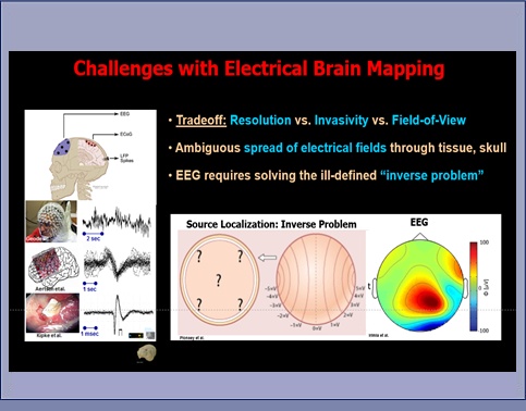
As part of the NIH Brain Initiative, we are developing acoustoelectric brain imaging (ABI)
as a new modality for noninvasive mapping of electrical current at higher spatial resolution than EEG.
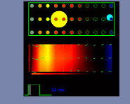
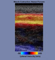
ABI exploits the acoustoelectric (AE) effect, an interaction between pressure (focused ultrasound) and tissue resistivity,
to map current densities in 4D (volume + time). By rapidly sweeping an ultrasound beam in the brain while recording the AE interaction signal,
it may be possible to image the human brain noninvasively, in real-time and at a higher resolution than standard EEG. As a new modality,
Transcranial Acoustoelectric Brain Imaging (tABI) would expand existing maps of brain connectivity,
refine their links to perception and behavior, and help repair lost function caused by a brain disease or injury.
|